
Posted by Admin / 25 January, 2022 /
Nowadays, there are hundreds of diseases. Some are quite fatal like metastatic cancers while others can be handled with proper care. It doesn't only refer to one organ system or two. Instead, the whole body feels the effects of such diseases. For example, corneal edema can put your patient's eye at risk.
So, the question is-Has technology helped us in corneal lesions?
The simple answer is yes. You have a slit beam imaging system to diagnose corneal lesions. Once you have interpreted the illness, eye care professionals can save the life of a patient.
Today, we will go through slit lamp illumination and understand various aspects like lighting techniques and the use of digital cameras.
Chapter 1: Slit Lamp imaging system basics
What is a slit lamp imaging system?
There are hundreds of methods to diagnose eye disorders. But, during all this, we need to understand what a slit lamp illumination can do for you?
A slit lamp imaging system is a device that is used to capture the images of the eyes. These are not simple digital camera images. Instead, high microscope magnification helps us to click focused and zoomed-in images.
Evolution of slit lamp biomicroscope
Slit-lamp biomicroscope has a long history. Let's go through different lighting techniques in the evolution of slit lamp biomicroscope.
First time in history, Purkinje used the first microscope in 1820 to examine the iris. Later on, several scientists and ophthalmologists tried the microscope.
However, as far as slit lamp illumination system is concerned, Allvar Gullstrand proposed the idea in 1911. In 1919, Vogt Henker made changes to the Gullstrand slit microscope and came up with better ideas.
In 1927, stereo cameras were used in slit lamps. In 1938, a new system allowed the movement along the horizontal axis.
1994 and onwards, we have digital options with contact lenses in our slit lamp biomicroscope.
What is the purpose of a slit lamp?
However, during all this, we have been studying all about the slit lamp illumination system. Can you guess the role of slit lamp illumination in our daily lives?
Why slit lamp lighting techniques are important to us?
Maybe you can't crack the code. But, no worries at all. I have listed the purposes of contact lenses and lighting techniques in the slit lamp illumination system.
- The ultimate goal to use this device in the optical section is to diagnose eye disorders.
- It can detect the pigmentary glaucoma
- You can analyze the cornea and retina.
There can be other purposes. But, one thing is common- it analyzes the eye structure and detects diseases important for the optical section.
What are parts of slit lamps?
There are multiple parts of the slit lamps that check the dilated pupil and eye diseases through the use of light beams.
Now, the question is - What parts does a slit lamp comprise?
No surprises at all. Instead, light beam like wide beam plays a crucial role. Based on location, there is an anterior chamber or anterior segment.
. Viewing Arm
The viewing arm has a microscope lens through which the patient can see. Here are three parts:
- Ocular lens
- Magnification dial
- Focus ring
. Illumination Arm
The illumination arm is quite important. It comprises major components such as a lens used to detect patient eyes. Here are three components of the illumination arm.
- Beam width
- Beam height
- Color of light- White, Cobalt blue, and green also called a red free filter.
You can adjust the beam width, height, and colors such as cobalt blue or red free.
. Patients positioning frame
Patients positioning frame helps patients to adjust their head position. Here are three major parts.
- Forehead strap
- Chinrest
- Height adjust
The patient positioning arm might be slightly nasal.
. Base
The base of the slit lamp is the basal part. It might have:
- Joystick-allows movements for separate adjustments.
- Light Brightness Knob
- Locking knob
How does the photo slit lamp system work?
Our eyes have small blood vessels. Apart from blood vessels, these might comprise parts like the iris, cornea, or retina. Whenever you visit your ophthalmologist, the first challenge is to detect the problem.
So, how does an ophthalmologist make a diagnosis?
Maybe he tries the light beam from the slit lamp. You need to look through the lens. With the help of a narrow beam or wide beam, the doctor will check which part is not functioning. For example, the optic nerve is not properly working. The image will not be being formed on the retina. So, the doctor will take further steps regarding treatment. Moreover, other parts like the iris and dilated pupil can be checked through the slit beam.
What are the methods of illumination in the slit lamp system?
There are multiple Slit-lamp illumination methods. I have listed some of them.
- Diffuse illumination
- Direct illumination
- Indirect illumination
- Retro illumination
- Tangential illumination
In diffuse illumination, you can find two other significant methods like the direct diffuse illumination and indirect diffuse illumination. Illumination is quite important for viewing path.
Chapter 2: How to install the slit lamp imaging module?
Before installation of the slit lamp imaging module, I would recommend checking the beam width and other beam light-related adjustments. If everything is fine, you can move ahead to install your phonto and capture images.
Step 1: Attach your phonto
Get your phonto ready. Phonto is a part that will attach to the anterior chamber of the instrument. First, remove your lenses from the attachment point.
Attach the phonto to the attachment point. Now, you can again attach the lenses to your their location. You know, what is the benefit of it? It will handle the phonto firmly and let you make slit beam adjustments effectively.
Step 2: Install your metal plate to your smartphone
The metal plate has two ends. One end will be adhesive to attach to the smartphone. Another end will stick to the phonto.
No matter you have iPhone or Android, it will work with both.
Step 3: Install your smartphone to the phonto
Have you connected the metal plate to your smartphone? If yes, just great. Now, you are good to go for the next step.
Install the smartphone to the phonto. And how will you do that? You have the metal plate for the adhesion. If your device is slack incorporated, you can setup it correctly to prevent any damage to your device.
Step 4: Use a camera or mobile App image system for images
Slit illumination will help you analyze the patient's eye and check whether there are chances for pigmentary glaucoma or not. Moreover, you can adjust the angle of the examination.
But, there is a question. And what question that is: How will you capture the images?
There are two ways to take pictures. First, you can use your phone camera to control and capture images with the required angle. Second is our image systems that control the image-taking options for you. So, you can use any way you like to take the images.
Chapter 3: Where can you buy the slit beam imaging module in China?
Finding an outstanding slit beam imaging module is always hard. You know, why? Because you need to conduct meticulous research. Just contrast options don't work out. Instead, you find a slit beam imaging module that is a perfect return for your investment.
So, is it possible to find a good module? Yes, I can help you find a better option. Here are our two models that will prove valuable to your eye care clinic.
Phonto
Phonto is a popular choice for the ophthalmologist when they want to capture the images.
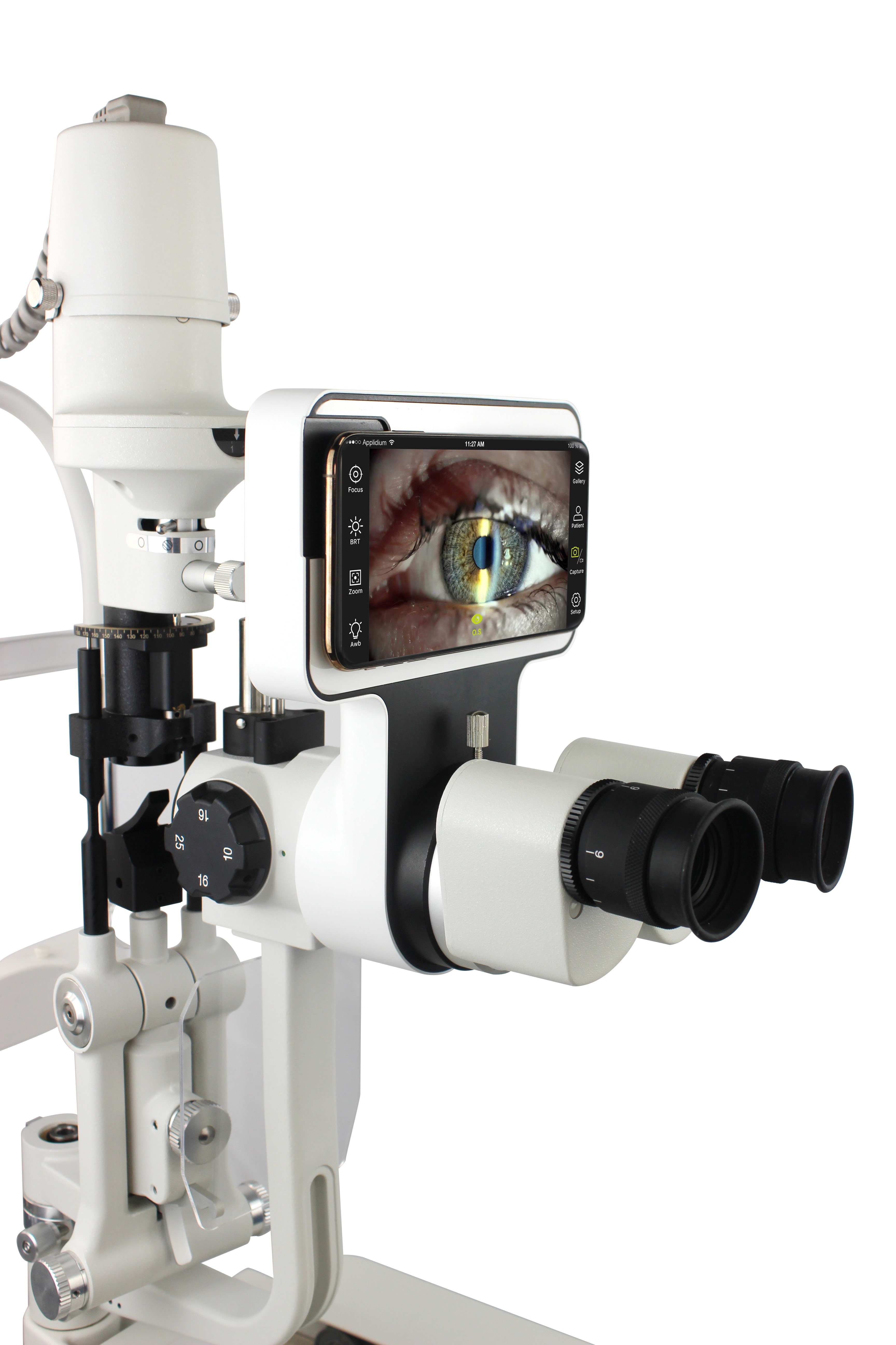
Here is why you should choose our slit lamp imaging module:
- It works with all the devices. No matter you use an Android device or iOS, it can work with both of them.
- Utilizes the phone camera to take high-quality images for detection of diseases in the cornea, iris, or retina. Pigmentary glaucoma can be easily detected with this.
- Available at a cheap price compared to our competitors with the quality you need.
- It is compatible with different models of slit lamps. For example, Topcon slit lamps or Zeiss lamps perfectly work with this.
Want to give it a try? Hit us a message right away.
Phoenix
Our Phoenix model of imaging module is a complete solution for the proper diagnosis of cornea, iris, and retina. It works with multiple models of imaging modules to help doctors achieve flexible options.
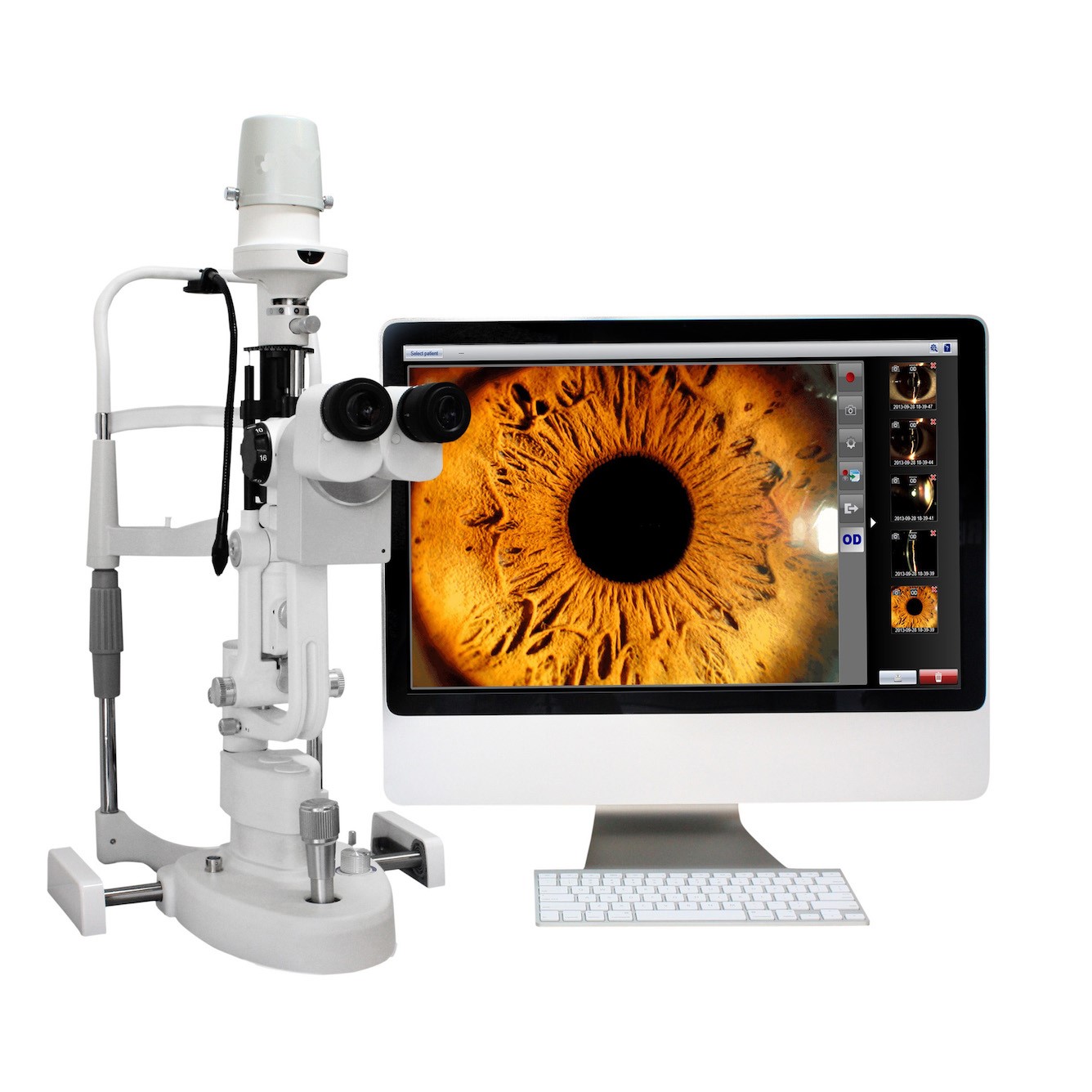
Here is what makes our Phoenix special:
- Takes high-quality images and videos in 1080p format.
- The power supply is 5 volts DC that is commonly available.
- Compatible with popular models of slit lamps available around the globe.
- Cheap option compared to competitors in this range
EndNote
There is one tip for you at the end. Always set up your own criteria to judge the instruments. If you are a doctor, you must learn about different components. Check the quality performance and find the cost-effective equipment. Not all slit lamp imaging modules are incredible. Instead, a few top-notch companies in China offer you reliable solutions for your medical care centers.